Say Goodbye to Stained Teeth with Porcelain Veneers
Patients often arrive after whitening has failed to improve tooth colour. For individuals considering dental veneers in Mississauga, ON, the main concern is usually persistent discoloration that has not responded to bleaching. In many of these situations, the colour change lies within dentin rather than on enamel. Because enamel allows light transmission, the underlying dentin shade directly influences visible appearance. Clinical examination includes radiographs to review bone height, inspection of the gingival tissue for inflammation, and vitality testing to confirm nerve health. If infection, active decay, or periodontal instability is identified, those conditions are managed before any enamel is reduced. Placing restorations on compromised tissue increases biological risk.
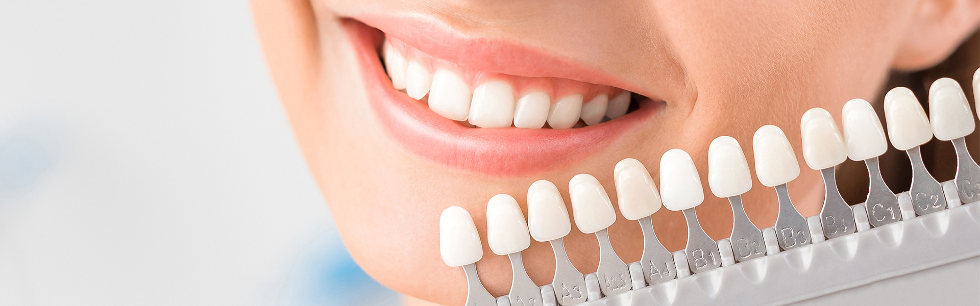
What Are Porcelain Veneers?
Porcelain veneers are thin ceramic restorations bonded to the front surface of teeth following controlled enamel reshaping. Preparation depth is measured to preserve sufficient enamel for adhesion, since bonding to dentin is less predictable. Enamel removal is permanent, so reduction is limited to what is required for space and contour.
The ceramic is fabricated with calibrated opacity to neutralize darker dentin tones. Occlusal contact patterns are analyzed before preparation. If anterior teeth absorb excessive functional force, stress may concentrate along the adhesive margin. In cases with heavy bite pressure or edge-to-edge contact, occlusal adjustment or protective stabilization may be recommended to reduce fracture potential.
Common Causes of Tooth Staining
Tooth discoloration develops from either external or internal sources. External stains attach to enamel through dietary pigments or tobacco exposure. Internal discoloration originates within dentin and may follow trauma, medication exposure, or pulpal degeneration.
Blunt trauma can disrupt blood flow inside the pulp chamber. As hemoglobin components degrade, internal staining may occur. Radiographic imaging helps identify root damage, bone changes, or periapical infection. If pulpal necrosis is diagnosed, endodontic treatment may precede cosmetic coverage. Treatment planning depends on pulp vitality, structural strength, and periodontal support.
How Porcelain Veneers Cover Stains
Veneers conceal discoloration by placing a ceramic layer over the prepared enamel surface. After reshaping the tooth, impressions or digital scans are obtained to coordinate laboratory fabrication. Ceramic thickness and internal shading are adjusted to block darker dentin without excessive bulk.
Adhesive bonding requires strict isolation. Saliva or blood contamination can interfere with resin polymerization and weaken the seal. Margins are inspected under magnification to confirm close adaptation to enamel and surrounding gingival tissue. Irregular margins may harbour plaque and contribute to inflammation or secondary decay. Follow-up visits allow observation of soft tissue response and occlusal stability over time.
Veneers vs. Teeth Whitening Treatments
Whitening systems function through peroxide diffusion into enamel, where oxidation alters stain molecules. Surface discoloration often responds. Deeper dentin staining shows limited improvement because penetration depth is restricted.
Veneers may be appropriate in circumstances such as:
- Intrinsic discoloration resistant to bleaching
- Enamel thinning that reduces whitening predictability
- Minor contour irregularities requiring reshaping
- Structural enamel defects affecting surface integrity
A cosmetic dentist in Mississauga will consult and compare enamel thickness, pulp condition, and occlusal loading before determining whether chemical whitening or restorative masking is more suitable. Recommendations are based on biological limitations rather than cosmetic preference alone.
Benefits of Choosing Porcelain Veneers
Bonded ceramic restorations distribute functional forces across the facial tooth surface when adequate enamel remains. Porcelain demonstrates lower surface porosity than enamel, reducing susceptibility to external staining.
Clinical considerations include:
- Refined contour that may support improved plaque control if margins remain smooth
- Colour stability because ceramic does not undergo metabolic change
- Structural reinforcement of weakened enamel areas when bonding conditions are favourable
Limitations must be discussed. Gingival recession may expose restoration edges. Parafunctional habits such as grinding can create repetitive stress and increase the risk of chipping or debonding. Periodic review is necessary to verify bond integrity and tissue health.
Who Is a Good Candidate for Porcelain Veneers?
Candidacy requires stable periodontal attachment, sufficient bone support, and absence of untreated infection. Inflamed gingiva must be treated and allowed to recover before enamel modification begins. Adhesion to compromised surfaces reduces long-term predictability.
Occlusal patterns are reviewed because lateral grinding forces may overload ceramic. In selected cases, fabrication of a protective night guard helps distribute stress. If tooth mobility or advanced bone loss is present, alternative treatment options may provide safer long-term outcomes.
Evaluation at a dental clinic in Mississauga includes radiographs, occlusal analysis, and detailed soft tissue examination before proceeding with irreversible preparation.
Final Thoughts
Veneers are considered when discoloration originates within dentin and cannot be predictably corrected with whitening agents. Enamel reshaping, adhesive bonding, and occlusal verification are deliberate procedural steps intended to maintain biological balance and structural integrity. Potential risks include postoperative sensitivity, marginal leakage, ceramic fracture under excessive load, and eventual replacement depending on wear patterns and periodontal changes.
At Pearl Dental Care, restorative decisions are based on radiographic evidence, periodontal findings, and functional analysis to support long-term oral stability.